SPECTRA NAVIGATION · MICROSURGERY

Spectra NavigationIntelligent Microsurgical Navigation System

Designed for deep-field visualization and quantitative guidance in microsurgery, the system integrates split-path stereoscopic imaging, miniature instrument tracking, and dynamic registration into one intraoperative platform.

Precision Visualization Intelligent Navigation Microscale Surgery
Spectra Navigation validation prototype
Split-path stereoscopic microscopic imaging systemValidation prototype

01 · Clinical Need

Microsurgery lacks a quantitative reference for deep anatomy

The relative positions of lesion boundaries, instrument tips, and critical anatomy are difficult to judge directly. As procedures move deeper and toward smaller structures, reliance on individual experience increases, along with training time and variability in risk.

Relevant procedure volume
2.25million/year

Estimated annual volume of core microsurgical procedures in China.

Clinical value of resection extent
+29.4months

Reported mean survival benefit of gross total versus subtotal resection for grade IV glioma.

Neurosurgeon density
0.78per 100,000 people

The supply of specialists cannot expand quickly, increasing the value of navigation for decision support and shorter learning curves.

Microsurgeon using an operating microscope
Deep structures are difficult to observe directly, making surgical decisions highly experience-dependentClinical setting
Surgical training setting
Training for complex procedures takes years, and regional capability gaps are difficult to close quicklyWorkforce bottleneck

Clinical and market figures should be published with their definitions, reference year, indication scope, and public sources.

02 · Limits of Existing Solutions

Millimeter-scale navigation creates both scale and workflow mismatches in the microscopic field

Conventional navigation is generally designed around rigid bony fixation, large reflective markers, and an open field. Microsurgery requires smaller tracking components, higher localization accuracy, and robustness to occlusion, glare, bleeding, and soft-tissue deformation.

Size comparison between a conventional navigation marker and microsurgical instruments
Conventional markers are poorly matched to the scale of microsurgical instrumentsScale comparison
The microscopic field changes the basic conditions for navigation

Existing systems typically depend on rigid anatomy, large markers, and one-time registration. In the microscopic field, instrument tips, working space, and recognizable features all become smaller while soft tissue continues to deform. Navigation must therefore maintain a trustworthy reference continuously rather than localize only once.

2–10 mm
Registration remains too coarse

The effective field and critical structures in microsurgery are often only millimeters or smaller, making conventional navigation error unsuitable for fine manipulation.

20–100 mm
Tracking markers are too large

Large reflective markers occupy working space, obstruct the view, and alter the feel and inertia of delicate instruments.

Dynamic soft tissue
The intraoperative environment keeps changing

A small field contains few usable features, while tissue deformation and fluid reflections can quickly invalidate a one-time registration.

03 · Product Concept

Turn what cannot be seen into actionable intraoperative measurements

The system provides depth, boundary, distance, and risk cues around the surgeon's active microscopic workflow, keeping preoperative imaging, instrument position, and the intraoperative view linked.

WITHOUT NAVIGATION · 01

Deep structures remain unseen and decisions depend on individual experience

  • Tissue boundaries lack quantitative references, making under-resection and over-resection difficult to assess
  • Surgeons must integrate multiple information sources themselves, increasing cognitive load
  • Training and supervision require extensive case experience
WITH SPECTRA · 02

Critical structures are quantified and updated with instrument position in real time

  • Displays the relative position of the instrument tip and lesion boundary
  • Overlays the preoperative model onto the intraoperative microscopic view
  • Provides a consistent reference for decisions, documentation, and review
Intraoperative microsurgery and navigation interface concept
The objective is concrete: bring boundary, distance, and risk cues into the current view without disrupting the surgeon's core operating habits.Product value still requires continued validation in real clinical workflows, against defined baselines, and through surgeon feedback.

04 · Core Technology

Three technologies address field visualization, instrument tracking, and continuous model alignment

Visualization reconstructs the 3D surgical field, precision tracking localizes the instrument tip, and intelligent registration updates the coordinate reference as tissue and the field change. These capabilities connect along a single navigation chain.

VISUALIZATION · 01

Visualization: split-path stereoscopic microscopic imaging

The three images show how visualization is implemented: coaxial visible and near-infrared acquisition, a validation prototype that establishes 3D metrology, and final integration with an operating microscope.

  • Visible and near-infrared light share one optical path for simultaneous field visualization and tracking-signal acquisition.
  • Stereo vision provides the 3D measurement foundation, converting microscopic images into depth and relative-position information.
  • Once integrated with the microscope, the system supplies stable observations for instrument tracking and dynamic registration.
Intelligent Registration
Instrument Tracking
High-precision Reconstruction
Coaxial optical-path diagram for split-path stereoscopic microscopy
01 · Coaxial split optical pathVisible + near-infrared
Split-path stereoscopic imaging validation prototype
02 · Optical validation prototype3D metrology foundation
Microscopic navigation prototype with integrated split-path stereo vision
03 · Microscope integrationSystem validation prototype

The microscope platform combines imaging and metrology, preserving the live surgical field while providing structural, depth, and relative-position references.

PRECISION · 02

Ultrafine optical fibers enable continuous instrument-tip tracking

Ultrafine optical fibers are embedded in microsurgical forceps, scissors, and related instruments to add active tip-tracking signals while preserving familiar dimensions, weight, and handling.

< 0.1 mmFiber diameter
0.009 mmR&D tracking accuracy
Dynamic demonstration of single-point mouse tracking
Single-point mouse trackingNavigation validation
Trackable microsurgical instrument with embedded ultrafine fibers
Trackable microsurgical instrumentStructural concept
Dynamic demonstration of fiber-instrument tracking and 3D reconstruction
Fiber tracking and 3D reconstructionDynamic validation

INTELLIGENCE · 03

Dynamic registration maintains a reliable reference at microscale

In robotic R&D validation, the system created an approximately 2 mm cranial window in mouse skulls averaging about 0.3 mm thick. The central capability is maintaining the preoperative plan, live view, and instrument position in one coordinate relationship to support high-precision operation.

74 casesRobot-assisted mouse craniotomy experiments
≈ 0.3 mmAverage mouse-skull thickness
≈ 2 mmCranial-window diameter
Dynamic adaptive registrationAlgorithm workflow
01
Establish initial alignment

Extract constraints from recognizable structures such as vessels and bone-surface texture to align the preoperative model with the intraoperative view.

02
Continuously evaluate deviation

Under a small field, occlusion, glare, and tissue change, the system continuously checks whether the current reference remains reliable.

03
Update coordinates online

Dynamic landmarks update relative positions and return the instrument trajectory, model, and live image to a common spatial relationship.

Robot performing a mouse craniotomy with navigation assistance
Robot-assisted mouse craniotomyHigh-precision operation demonstration
Automated registration and calibrationLive process
Close-up of a mouse cranial window
Cranial-window close-upApproximately 2 mm
Scale reference for a small cranial-window model
Small cranial-window modelScale reference
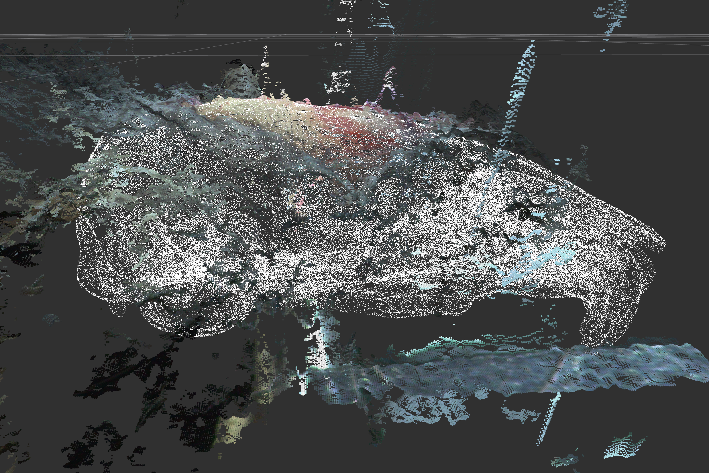
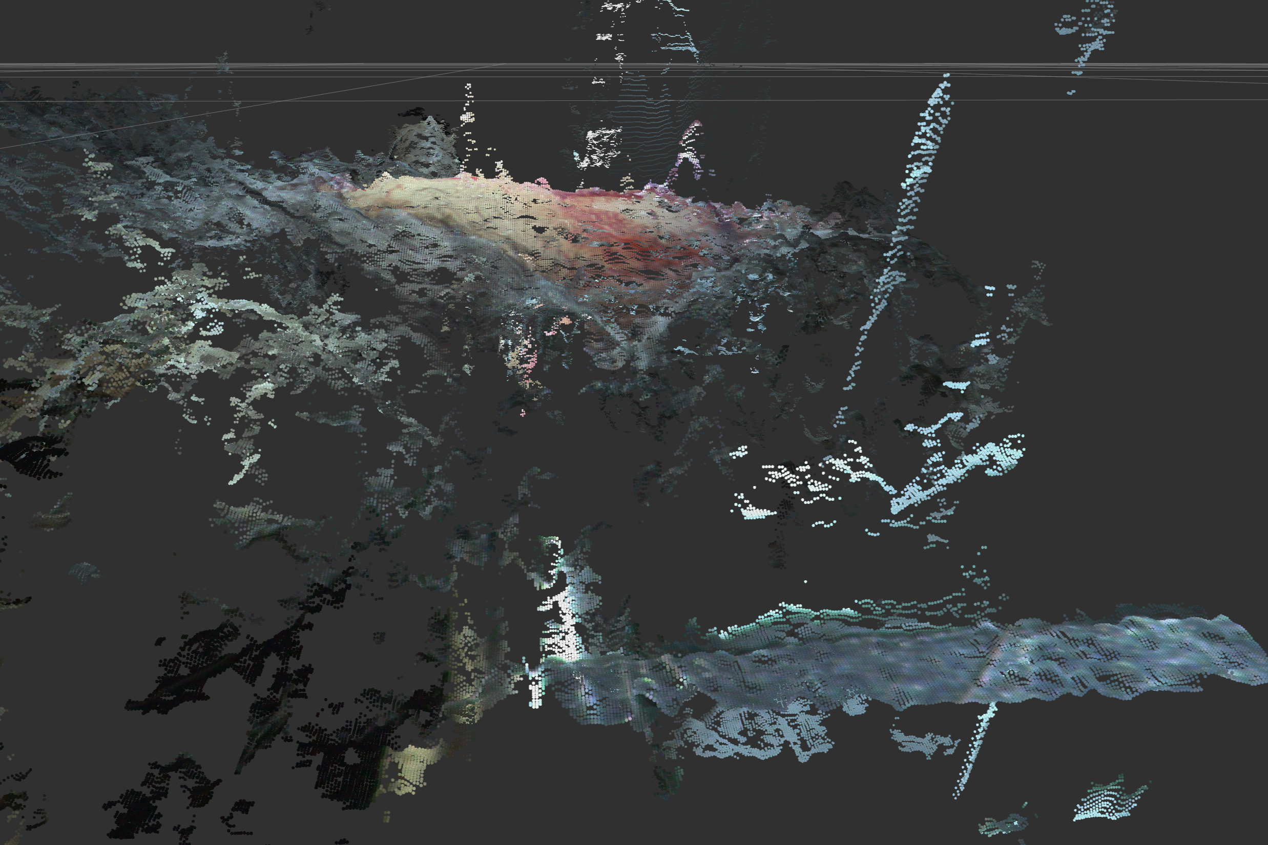
Mouse-head point cloud and coordinate reconstruction
Point cloud and spatial coordinates3D reconstruction

These results demonstrate R&D-stage system capabilities. Medical use, performance claims, and clinical approval status remain subject to subsequent validation and public regulatory documents.

05 · Product System

The product is an integrated intraoperative hardware-software system

The system combines a 3D navigation workstation, split-path stereo operating microscope, and trackable fiber-optic instruments. Dynamic registration, AI recognition, and intraoperative visualization connect all three components.

Split-path stereo operating microscope
02 · Split-path stereo operating microscopeStereoscopic field and depth measurement
3D navigation workstation
01 · 3D navigation workstationPlanning, localization, and model linkage
Trackable fiber-optic instruments
03 · Trackable fiber-optic instrumentsContinuous instrument-tip tracking
Complete navigation systemDynamic registration · Intraoperative visualization · Instrument tracking
01
3D navigation workstation

Integrates preoperative imaging and 3D models for planning, intraoperative localization, model linkage, workflow recording, and postoperative assessment.

02
Split-path stereo operating microscope

Acquires visible and near-infrared signals coaxially, providing a stereoscopic field, depth measurement, and observations for instrument tracking and dynamic registration.

03
Trackable fiber-optic instruments

Embedded ultrafine fibers continuously localize the instrument tip and calculate its relative position to lesion boundaries and critical structures in real time.

06 · Development and Intellectual Property

Development is progressing from prototype validation toward registration preparation

The product path includes research prototypes, animal studies, registration prototypes, clinical trials, and NMPA review. Timing remains dependent on experimental progress, regulatory consultation, and funding.

2021
Project R&D

Completed method innovation and exploration of the core technical route.

Completed
2023
First-generation prototype

Advanced human-machine interaction validation and prototype development.

Completed
2026
Product development and animal studies

Continuing iterations of the tracking system, navigation software, and product engineering.

In progress
2027
Registration prototype and clinical-trial application

Prepare registration documentation and advance acceptance and clinical trials.

Planned
2028 Q3
NMPA Class III medical-device review

This target depends on regulatory consultation and trial progress; the final timeline will reflect actual development.

Target
Patent certificate and application document 1 Patent certificate and application document 2 Patent certificate and application document 3

IP PORTFOLIO

The team has filed or secured nine patents covering navigation, registration, instrument tracking, and surgical robotics

The Japanese application for a navigation system, algorithm, and instruments, together with the Chinese application for a 3D calibration board and performance-evaluation method for stereo microscopic imaging, are most closely aligned with the current product. The remaining patents reflect prior technical work; ownership, licensing scope, and freedom to operate require item-by-item confirmation during investment and partnership due diligence.

View patent list
  1. Japan: Navigation system, algorithm, and instruments, 2025-284200, pending
  2. China: 3D calibration board and performance-evaluation method for stereo microscopic imaging systems, CN202610805057.1, pending
  3. China: Coaxial guidewire-catheter drive device and control method for endovascular intervention, CN114177480A, granted
  4. China: Robotic puncture-positioning device for biliary intervention, CN215874870U, granted
  5. China: Robot and equipment for endovascular intervention, CN110200700B, granted
  6. WIPO:FORCE FEEDBACK APPARATUS, AND APPLICATION THEREOF,WO2022204869A1,Granted
  7. China: Force-feedback apparatus and applications, CN113081275A, granted
  8. China: Radioactive seed implantation robot, CN110548218A, granted
  9. China: Portal-vein radioactive seed stent implantation mechanism, CN305545928S, granted

07 · Market and Commercial Path

Enter through modules and software, then progress toward a proprietary complete system

Installed systems establish the scale base, while software licensing and dedicated consumables create recurring revenue. An early OEM route can reduce upfront investment in full-system development, channels, and registration infrastructure.

GLOBAL SURGICAL NAVIGATION

83.8 → 184.7USD 100M

$10.09B projected increase over six years

The global market is projected to grow from $8.38B in 2024 to $18.47B in 2030, approximately 2.2 times its current size, at an estimated 14.0% CAGR.

CHINA SURGICAL NAVIGATION

2.5 → 5.9USD 100M

$340M projected increase over six years

China's market is projected to grow from $250M in 2024 to $590M in 2030, approximately 2.4 times its current size, at an estimated 15.3% CAGR.

PHASE 01 · OEM

Module supply + software licensing

  • Split-path stereo-vision hardware moduleRMB 0.8–1.2M/system
  • Software licenseRMB 0.2–0.3M/system/year
  • Dedicated consumablesRMB 500–1,500/case
Approximately RMB 25M/yearIllustrative case: 100 installed systems and 100% consumables adoption.
PHASE 02 · OWN BRAND

Proprietary complete-system sales

  • Complete-system list priceRMB 2.0–2.5M/system
  • Enhanced software licenseRMB 0.3–0.5M/system/year
  • Dedicated consumablesRMB 500–1,500/case
Approximately RMB 35M/yearIllustrative case: 100 installed systems and 100% consumables adoption.

Market size, pricing, and revenue examples illustrate the commercial logic. Actual performance will depend on source data, installation ramp, gross margin, consumables frequency, channel discounts, and the post-registration launch schedule.

08 · Competitive Landscape

The product targets the intersection of microscopic-field fit, dynamic registration, and instrument tracking

Existing solutions include bone-fixed navigation, non-invasive navigation, and vision-enhanced microscopes. Spectra Navigation focuses on microscopic-scale compatibility and compensation for soft-tissue deformation.

Product / CountryProductRegistration methodMicroscopic-field fitSoft-tissue compensationClosed-loop capabilityStandard indications
Proprio Paradigm
(United States)
Proprio Paradigm product appearanceBone-fixedNot compatibleYesRegistration + trackingSpine
Brainlab Microscope Navigation
(Germany)
Brainlab Microscope Navigation product appearanceBone-fixedNot compatibleNoRegistration + trackingGeneral orthopedics or spine
Medtronic StealthStation S8
(United States / Ireland)
Medtronic StealthStation S8 product appearanceBone-fixedNot compatibleNoRegistration + trackingGeneral neurosurgery / general orthopedics
Beyeonics One
(Israel, operating microscope)
Beyeonics One product appearanceNo registrationCompatibleNoVisualization enhancement onlyGeneral ophthalmology
Huake Precision
(China)
Huake Precision navigation productBone-fixedNot compatibleNoRegistration + trackingGeneral neurosurgery / general orthopedics
SeaSpine / Orthofix 7D FLASH
(United States)
SeaSpine Orthofix 7D FLASH product appearanceNon-invasive fixationNot compatibleNoRegistration + trackingNeurosurgery (brain tumors)
Spectra Navigation Intelligent Microsurgical Navigation System
(This product)
Spectra Navigation microsurgical navigation system conceptNon-invasive fixationCompatibleYesRegistration + trackingGeneral neurosurgery / ENT / ophthalmology / reconstructive surgery

Competitor specifications and approval status change over time. Current specifications, indications, registration status, and soft-tissue compensation capabilities should be verified item by item before public release.

09 · Team

A team spanning clinical practice, optics, algorithms, automation, and commercialization

Core members have research and academic experience at the University of Tokyo, Tsinghua University, Harvard University, Yale University, and UNSW, with R&D backgrounds in surgical robotics, medical imaging, and navigation systems.

Founder and CEO Dr. Xiaofeng Lin

FOUNDER & CEO

Dr. Xiaofeng Lin

Experience

  • Project Researcher in medical devices, Faculty of Medicine, the University of Tokyo
  • PhD in biomedical engineering, the University of Tokyo
  • Master's degree, Shenzhen Institutes of Advanced Technology, University of Chinese Academy of Sciences
  • Former co-founder of Hengle Medical Technology, a Series B+ company

Achievements

  • Global Medical Robotics Innovation Design Competition: Silver Award
  • 6th China International Internet+ College Student Innovation and Entrepreneurship Competition: Gold Award
  • IROS Workshop Best Poster Award in surgical robotics
  • Multiple publications in leading journals and conferences on surgical navigation
  • Eight patents in surgical robotics and surgical navigation
Team member Enduo Zhao

Enduo Zhao

Automation

  • Postdoctoral researcher in biomedical engineering, Tsinghua University
  • PhD in mechanical engineering, the University of Tokyo
  • Seven journal and conference papers on surgical robotic systems
  • Multiple robotics patents
Team member Yuhan Song

Yuhan Song

Computer Vision

  • PhD candidate in biomedical engineering, the University of Tokyo
  • Project Researcher at a Japanese national university, leading algorithm development and engineering implementation for robotic medical systems
  • JSPS Research Fellow working on microsurgical navigation
  • Four journal and conference papers in medical image processing
Team member Shenghao Jiang

Shenghao Jiang

Clinical Research

  • MD candidate, UNSW Sydney
  • Master's degree in computer science and engineering, Harvard University
  • Several years of R&D experience in AI vision algorithms for autonomous navigation
  • Eight journal and conference papers in medicine and robotics
Team member Zenghui Yu

Zenghui Yu

Market & Operations

  • Several years as a manufacturing-focused consulting analyst at an investment firm
  • Several years leading overseas business development at a Japanese manufacturer
  • Master's degree in commerce, Komazawa University
Team member Guanhao Lin

Guanhao Lin

Technology Transfer & Finance

  • AI consultant and systems engineer at an AI systems-integration consultancy
  • Several years as a consulting associate in venture capital
  • Master's degree in systems innovation, the University of Tokyo
Team member Gina Quan

Gina Quan

Technology Transfer & Partnerships

  • PhD candidate, Faculty of Medicine, the University of Tokyo
  • Master's degree in biomedical engineering, Yale University
  • Former hardware and software developer at a medical robotics company
Chief Advisor Jian Lu

CHIEF ADVISOR

Jian Lu

Appointments

  • Associate Chief Physician, Young Chief Professor, and PhD supervisor at Zhongda Hospital, Southeast University
  • Young Chang Jiang Scholar
  • Director of the Talent Office, Zhongda Hospital, Southeast University
  • Deputy Director of Interventional and Vascular Surgery, Zhongda Hospital, Southeast University
  • Vice Chair, Interstitial Tumor Implantation Committee, Chinese Anti-Cancer Association
  • Member, Youth Committee of the Interventional Physicians Branch, Chinese Medical Doctor Association

Achievements

  • Principal investigator for ten provincial- and ministerial-level projects, including the Jiangsu Distinguished Young Scholars Fund, a National Key R&D Program subproject, and National Natural Science Foundation grants
  • More than 50 SCI papers as first or corresponding author, including work in Lancet Gastroenterology & Hepatology (cover), Nature Communications, and Journal of Hepatology
  • Sixteen granted or pending patents
  • First Prize, 2022 Jiangsu Science and Technology Award
  • Young Investigator awards from leading interventional societies in North America, Europe, and Asia-Pacific
  • ASCO GI Conquer Cancer Merit Award
  • Outstanding Young Scholar, Interventional Physicians Branch of the Chinese Medical Doctor Association
  • National Distinguished Physician: Emerging Young Talent

10 · Risks and Current Needs

The next priority is converting technical performance into clinical evidence and a registrable product

The project remains in R&D and validation. Clinical workflow, funding, procurement logic, and the regulatory path must converge in parallel; delay in any one area will affect productization.

RISK 01

Clinical workflow validation

Real-world workflows and clinical-value endpoints still require validation, and the product definition may undergo several iterations.

Response: prioritize brain-tumor resection and conduct joint validation with leading tertiary hospitals to establish workflows and comparison baselines.
RISK 02

Cash flow and financing window

Early hardware integration requires concentrated investment, while animal studies, prototypes, and registration preparation create continuous funding needs.

Response: use a modular OEM path and combine project funding with collaborative research to reduce upfront investment.
RISK 03

Procurement logic for a new market

The paying entity, departmental budget, and ROI evidence must be established progressively through demonstration cases and procurement discussions.

Response: first build a clinical evidence package, then validate pricing, channels, and the pace of indication expansion.

RESOURCE PRIORITIES

Three resource groups must be secured in parallel

Clinical validationAnimal studies, close clinician collaboration, data collection, and staged reviews.
Product engineeringHardware and software optimization, systems integration, industrial design, and prototype iteration.
Operations and complianceIntellectual-property strategy, regulatory planning, quality systems, manufacturing, and supply-chain management.
Key hires and external advisorsClinical program leads, hardware and software engineers, quality and regulatory staff, plus advisors for animal studies, legal matters, and manufacturing scale-up.

FUND ALLOCATION

Use-of-funds framework

Clinical validation
30%
Product engineering
30%
Regulatory and quality
20%
Operations
15%
Contingency
5%

The financing requirement will be calculated against an 18–24 month milestone budget, cash flow, and registration plan.

SPECTRA NAVIGATION

Give microsurgery a quantitative intraoperative reference

Spectra Navigation aims to bring navigation into smaller, deeper, and more dynamic microscopic fields. The next stage requires clinical, industry, and investment partners to advance validation, registration, and scalable deployment.

CLINICALJoint validation and clinical workflow co-development
INDUSTRYCollaboration across microscopes, instruments, and medical devices
CAPITALSupport prototype iteration, animal studies, and registration preparation